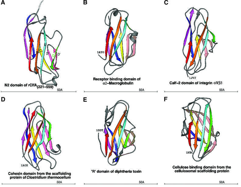
Fig. 5. Ribbon diagram of crystal structures with the DEv-IgG fold colored in rainbow fashion as in Figures 2 and 4. (A) N2 domain of ClfA. (B) Receptor-binding domain of α2-macroglobulin (PDB code: 1AYO, Z-score: 8.7, r.m.s.d.: 2.8, equiv. resid: 133). (C) Calf-2 domain of extracellular segment of the integrin αVβ3 (PDB Code: 1JV2, Z-score: 8.4, r.m.s.d.: 12.3, equiv. resid: 149). (D) Cohesin domain from the scaffolding protein of Clostridium thermocellum (PDB Code: 1AOH, Z-score: 7.6, r.m.s.d.: 3.4, equiv. resid: 123). (E) The R-domain of diphtheria toxin (PDB Code: 1DDT, Z-score: 7.2, r.m.s.d.: 4.2, equiv. resid: 129). (F) The cellulosomal scaffolding protein (PDB Code: 1NBC, Z-score: 7.0, r.m.s.d.: 3.0, equiv. resid: 109). Z-scores, r.m.s.d. values and equiv. resid. are from DALI.

An official website of the United States government
Here's how you know
Official websites use .gov
A
.gov website belongs to an official
government organization in the United States.
Secure .gov websites use HTTPS
A lock (
) or https:// means you've safely
connected to the .gov website. Share sensitive
information only on official, secure websites.
