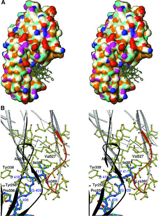
Fig. 6. (A) Stereo surface plot showing seven solutions of the Fg γ-chain peptide docked into a hydrophobic pocket between N2 and N3 domains. The hydrophobic, polar, positive and negative residues are shown in white, magenta, blue and red, respectively. Cyan and gold represent hydrogen bond donors and acceptors, respectively. (B) Stereo ribbon diagram showing the interactions of residues 408AGDV411 (blue) of the γ-chain of Fg with residues from both the N2 (dark gray) and N3 (white) domains of rClfA(221–559). The ribbon of the glycine-rich region 532GSGSGDGI539 is colored in orange.

An official website of the United States government
Here's how you know
Official websites use .gov
A
.gov website belongs to an official
government organization in the United States.
Secure .gov websites use HTTPS
A lock (
) or https:// means you've safely
connected to the .gov website. Share sensitive
information only on official, secure websites.
