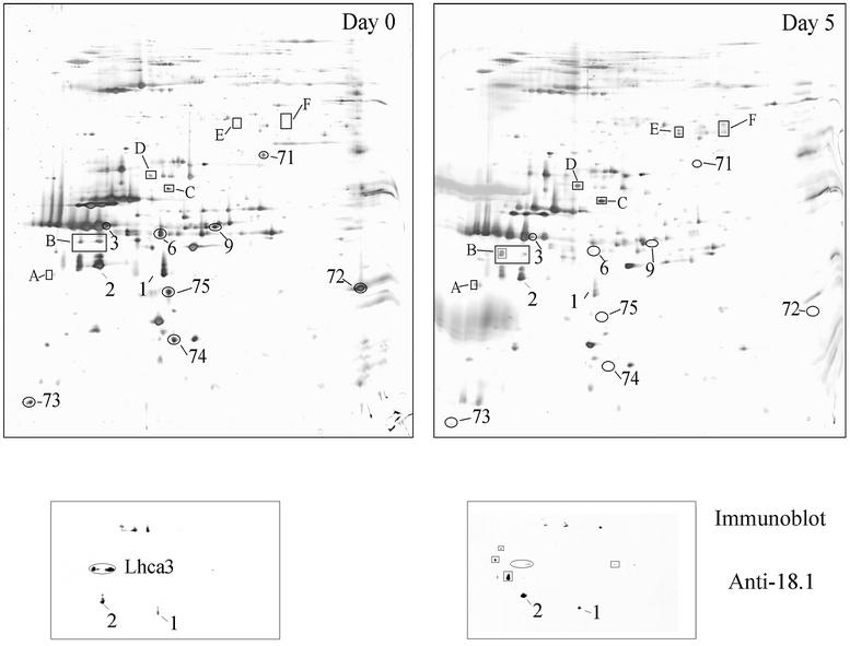
Fig. 5. (A) The top two panels (0 day and 5 day) represent silver-stained 2-D gels of thylakoid membranes from wild-type cells before and after 5 days of growth in Fe-deficient (0 µM Fe) medium, respectively. The labeled spots have been identified previously as follows: spots 1 and 2 correspond to the inner LHCI subunits Lhca1 and presumably Lhca4, respectively, while the labeled spots correspond to isoforms of Lhca3. (B) Immunodetection of LHCI subunits in thylakoid membranes separated by 2-DE as shown in (A), using an anti-serum (18.1; Bassi et al., 1992) which recognizes epitopes that are common to most of the LHCI subunits.

An official website of the United States government
Here's how you know
Official websites use .gov
A
.gov website belongs to an official
government organization in the United States.
Secure .gov websites use HTTPS
A lock (
) or https:// means you've safely
connected to the .gov website. Share sensitive
information only on official, secure websites.
