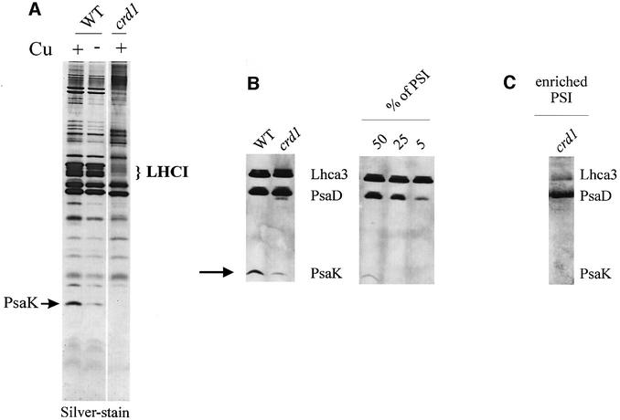
Fig. 9. (A) Silver-stained gel (Schägger and von Jagow, 1987) of enriched PSI/LHCI particles from +Cu (6 µM Cu) and –Cu (0 µM added Cu) wild-type and +Cu crd1 mutant cells. The migration of the PSI-K subunit is indicated with an arrow. (B) Immunoblot analysis to compare the abundance of PSI-D, Lhca3 and PSI-K in thylakoid membranes from wild-type and crd1 cells grown in TAP medium with normal Fe and Cu. The migration of PSI-K is indicated with an arrow. To determine the amount of PSI-K that is still present with the crd1 thylakoids, we performed in parallel dilution series where the total amount of thylakoid membrane protein loaded remained the same but the amount of PSI was diminished by mixing wild-type thylakoids with thylakoids isolated from a PSI-deficient mutant. The corresponding immunoblot shows that PSI-D and PSI-K signals vanish as the amount of PSI decreases. From this experiment, we can estimate that the amount of PSI-K in crd1 thylakoids is diminished to ∼50% of the quantity found in wild type. (C) Immunodetection of PSI-D, Lhca3 and PSI-K in enriched PSI/LHCI particles from +Cu crd1 cells.

An official website of the United States government
Here's how you know
Official websites use .gov
A
.gov website belongs to an official
government organization in the United States.
Secure .gov websites use HTTPS
A lock (
) or https:// means you've safely
connected to the .gov website. Share sensitive
information only on official, secure websites.
