
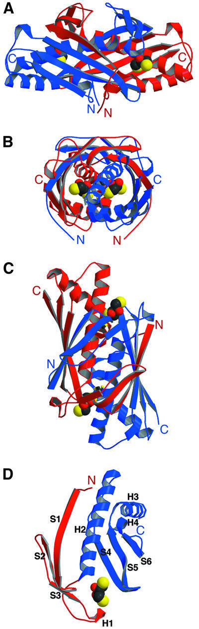
Fig. 2. (A–C) Structure of the Ohr dimer. One monomer is in red, the other is in blue. Three 90° views of the Ohr dimer bound to DTT. (D) Structure of the Ohr monomer depicting the two domains (the N-terminal subdomain is shown in red and the C-terminal subdomain is shown in blue) and a DTT molecule. DTT is shown in CPK format, with oxygen atoms in red, carbon atoms in black and sulfur atoms in yellow.