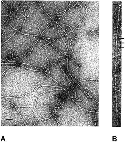
Fig. 1. Electron micrographs of negatively stained ParM filaments. (A) Typical overview of a grid containing ParM filaments. The protein (1 mg/ml) was incubated in polymerization buffer (30 mM Tris–HCl pH 7.5, 100 mM KCl, 2 mM MgCl2, 1 mM DTT) in the presence of 2 mM ATPγS for 5 min at room temperature. Scale bar, 100 nm. (B) ParM filament at higher magnification, showing the helical arrangement of the protofilaments. The crossovers are indicated by arrows. Scale bar, 100 nm.

An official website of the United States government
Here's how you know
Official websites use .gov
A
.gov website belongs to an official
government organization in the United States.
Secure .gov websites use HTTPS
A lock (
) or https:// means you've safely
connected to the .gov website. Share sensitive
information only on official, secure websites.
