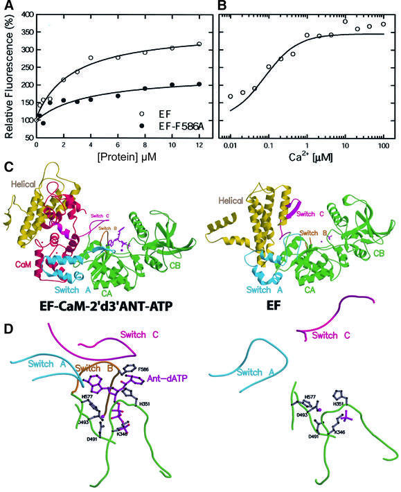
Fig. 2. The binding of 2′d3′ANT-ATP to EF. (A) Equilibrium titration of 2′d3′ANT-ATP–CaM with EF and EF-F586A. (B) Calcium titration of fluorescence enhancement by EF–CaM. 2′d3′ANT-ATP was added to a final concentration of 0.5 µM and the indicated free calcium concentrations were achieved by buffering with 10 mM EGTA. λexc = 320 nm and the optimal fluorescence emission of EF–CaM–2′d3′ANT-ATP (412 nm) was normalized to give the fold of enhancement. (C) Secondary structure of EF–CaM–2′d3′ANT-ATP in comparison with EF alone. (D) The active site of EF in the presence and absence of CaM and 2′d3′ANT-ATP.

An official website of the United States government
Here's how you know
Official websites use .gov
A
.gov website belongs to an official
government organization in the United States.
Secure .gov websites use HTTPS
A lock (
) or https:// means you've safely
connected to the .gov website. Share sensitive
information only on official, secure websites.
