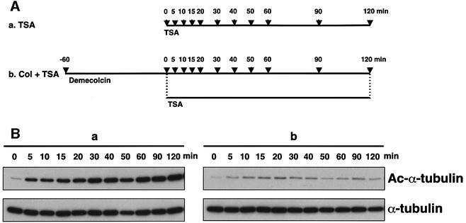
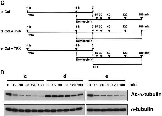
Fig. 7. Acetylation dependent on tubulin polymerization. (A) Schematic representation of experimental procedures for tubulin acetylation. Bars indicate the periods during which cells were treated with the drugs. Arrows indicate the time-points at which cells were taken for immunoblot analysis (B). (B) Effect of demecolcin pretreatment on TSA-induced tubulin acetylation. The amounts of acetylated and total tubulin in the cells in the time-course experiments designed as in (A) were determined by immunoblotting with anti-acetylated α-tubulin (upper) and anti-α-tubulin (lower) antibodies. (C) Schematic representation of experimental procedures for tubulin deacetylation. Bars indicate the periods during which the cells were treated with drugs. Arrows indicate the time-points at which cells were taken for immunoblot analysis (D). (D) Deacetylation of depolymerized tubulin. The amounts of acetylated and total tubulin in the cells treated with various drugs in the time-course experiments designed as in (C) were determined by immunoblotting with anti-acetylated α-tubulin (upper) and anti-α-tubulin (lower) antibodies.

An official website of the United States government
Here's how you know
Official websites use .gov
A
.gov website belongs to an official
government organization in the United States.
Secure .gov websites use HTTPS
A lock (
) or https:// means you've safely
connected to the .gov website. Share sensitive
information only on official, secure websites.

