
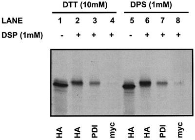
Fig. 4. Reduced and oxidized PDI associates with the S3 C-propeptide. The S3 C-propeptide was translated in a rabbit reticulocyte lysate in the presence of SP cells. SP cells were isolated by centrifugation, and then treated with DTT (lanes 1–4) or DPS (lanes 5–8). Excess DTT and DPS was removed by centrifugation before samples were chemi cally cross-linked with DSP or left untreated. Cross-linked and non-cross-linked samples were immunoprecipitated using the indicated antibodies before separation by SDS–PAGE on a 10% polyacrylamide gel, and translation products were visualized by autoradiography.