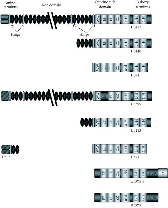
Figure 1.
Dystrophin and the dystrophin-related proteins specific to brain. Schematic representation of dystrophin and the dystrophin-related proteins, and the similarities shared by each protein. Shown are the four distinct regions of dystrophin: the amino-terminus, the rod-domain interrupted by the four proline-rich hinge regions, the cysteine-rich domain, and the carboxy-terminus. Abbreviations used: Hinge: hinge regions, Cysteine: cysteine-rich domain, WW: PPxY-binding WW motif, EF: Ca2+-dependent twin EF-hand motifs, ZZ: Zn2+-dependent zinc fingers, α-H: α-helical domain, L-H helical leucine heptads which makeup the coiled-coil domain, DUR: dystrobrevin-unique region, Y: region of tyrosine phosphorylation.

