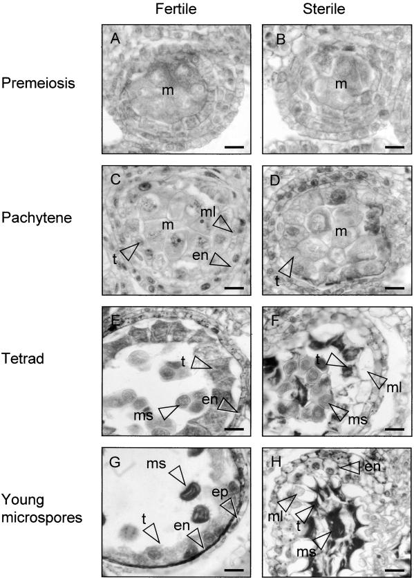
Figure 1.
Anther Development in Florets of the Fertile and Sterile Sunflower Lines.
Four stages of anther development in the fertile line and the corresponding stages of development in the sterile line were compared using histological staining. The images are of cross-sections through single locules.
(A) Fertile line, premeiosis.
(B) Sterile line, premeiosis.
(C) Fertile line, pachytene.
(D) Sterile line, pachytene.
(E) Fertile line, tetrad stage.
(F) Sterile line, tetrad stage.
(G) Fertile line, stage 6 according to Horner (1977): young microspores.
(H) Sterile line, stage 6 as in (G).
en, endothecium cell(s); ep, epidermal cell(s); m, meiocytes; ml, middle layer; ms, microspore(s); t, tapetal cell(s). Bars = 20 μm.

