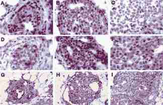
Figure 3.

Immunohistochemical staining for (a-c) cyclin D1, (d-f) p16INK4a(g-I) bromodeoxyuridine. Positive staining cells appear brown, counter-stained negative cells appear purple. (a), (d), and (g) are serial sections of an intraductal proliferation (IDP) from a Copenhagen rat; (b), (e), and (h) are serial sections of a large IDP from a Wistar-Furth rat; and (c), (f), and (I) are serial sections of a tumor from a Wistar-Furth rat. Note the overexpression of cyclin D1 in Wistar-Furth lesions (b and c) but not in the Copenhagen IDP (a). All lesions are from mammary glands of rats 37 days after MNU treatment. (a-f) 1000× magnification and (g-I) 400× magnification.
