Figure 6.
Expression of the DAD1 Gene.
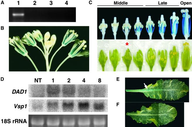
(A) RT-PCR analysis of DAD1 expression using total RNA isolated from Arabidopsis organs. Lane 1, wild-type flower bud clusters; lane 2, dad1 flower bud clusters; lane 3, wild-type leaves; lane 4, wild-type roots.
(B) and (C) Histochemical localization of GUS activity in DAD1::GUS plants. (B) An inflorescence. Blue signals are observed only in the stamen filaments. (C) A series of flower buds on an inflorescence. Detached flower buds were photographed (bottom row) and stained with X-Gluc solution (see Methods) after removing the sepals and petals (top row). The developmental stages of flower buds are shown above. The asterisk indicates the flower bud whose anther has begun to turn yellow.
(D) Wound-induced expression of the DAD1 gene. Gel blot analysis was performed using 10 μg of total RNA from rosette leaves harvested at the indicated times (hours) after wounding. NT, not treated. Top, RNA gel blots probed with DAD1 cDNA; middle, the same blots probed with Arabidopsis Vsp1 cDNA; bottom, ethidium bromide staining of 18S rRNA bands to confirm equal loading.
(E) and (F) Wound induction of GUS activity in rosette leaves of DAD1::GUS plants. (E) A leaf harvested and stained 1 hr after wounding showing GUS activity mainly in the midvein (arrow). (F) A leaf harvested from unwounded control plants showing no GUS activity.

