Figure 1.
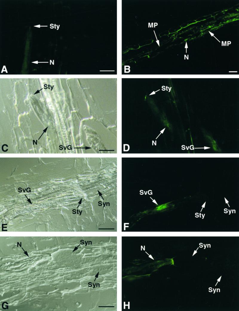
Immunolocalization of TCN EGases during Parasitism of Tobacco Roots.
(A) Longitudinal section through a tobacco root 24 hr after inoculation, with J2 of TCN (N) probed with mouse preimmune serum.
(B) Longitudinal section through a tobacco root 24 hr after inoculation, with TCN J2 probed with GR-ENG-1 antiserum. Binding of the GR-ENG-1 antiserum (green fluorescence) is observed along the migratory path (MP) of multiple migratory juveniles within the root cortical tissue. An arrow points to a nematode tail (N).
(C) Bright-field view of a longitudinal section through a tobacco root 24 hr after inoculation with J2 of TCN (N).
(D) Same section as (C) showing binding of GR-ENG-1 antiserum on the cell wall just outside the head of the nematode and within the nematode's subventral secretory gland cells (SvG).
(E) Longitudinal section through a tobacco root containing a sedentary parasitic J2 of TCN during early syncytium (Syn) development.
(F) Same section as (E) showing specific binding of GR-ENG-1 antiserum to the subventral secretory gland cells but not within the developing syncytium.
(G) Section through a tobacco root containing a sedentary parasitic J2 feeding from a well-developed syncytium.
(H) Same section as (G) showing slight binding of GR-ENG-1 antiserum to the surface of the nematode but not within the syncytium. Autofluorescence of plant cell wall adjacent to mematode head is visible.
Sty, nematode stylet. Bars = 50 μm.

