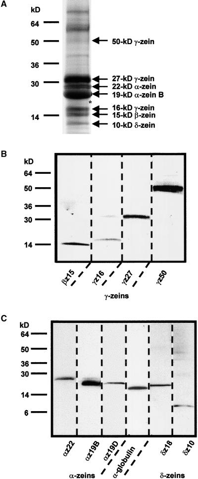
Figure 6.
Immunodetection of Proteins in Maize Endosperm Extracts.
(A) Total protein from B73 endosperm was separated by 4 to 20% gradient SDS-PAGE and stained with Coomassie blue. Positions of molecular mass markers are indicated at left. Proteins that were clearly identified with monospecific polyclonal antibodies are specified at right. The asterisk indicates the approximate position of the 19-kD α-zein D, the 18-kD δ-zein, and the 18-kD α-globulin polypeptides, all of which migrated to a similar position.
(B) and (C) Immunoblots of the protein extract in (A) separated by 4 to 20% gradient SDS-PAGE (B) and by 10 to 20% SDS-PAGE (C). Identical lanes were cut from the blots and incubated with the designated monospecific antibody (for abbreviations, see Table 1).

