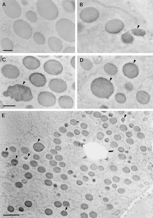
Figure 9.
Immunodetection of the 50-kD γ-Zein and the 18-kD α-Globulin in Protein Bodies of 20-DAP Endosperm.
Protein bodies were labeled singly with 50-kD γ-zein antibodies (A) or 18-kD α-globulin antibodies (B) and double labeled with 50-kD γ-zein antibodies (10-nm gold beads) and 19-kD α-zein antibodies (5-nm gold beads) (C) or 18-kD α-globulin antibodies (10-nm gold beads) and 19-kD α-zein antibodies (5-nm gold beads) (D). (E) shows a representative transmission electron micrograph showing a region of the third to fifth starchy endosperm cell layers labeled with 18-kD α-globulin antibodies (10-nm gold beads) and 19-kD α-zein antibodies (5-nm gold beads). The large empty hole is the former location of a starch grain. α-Globulin–containing protein bodies are indicated by arrowheads in (B), (C), (D), and (E). Bars = 0.4 μm in (A) and (C) and 2 μm in (E).

