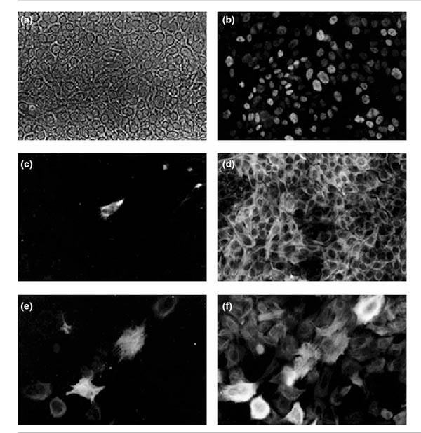
Figure 5.

Immunocytochemical analysis of KIM-2 cells cultures at 37°C. (a) Phase contrast. (b) The same field showing nuclear staining with a T-Ag-specific antibody (Pab 419). (c) Staining with a vimentin monoclonal antibody (Vim-13.2) was detected in some fields. (d) Strong positive staining with a keratin 18-specific monoclonal antibody (LE61), a luminal epithelial marker. Scale bar for (a)-(d) 50 μm. (e) Staining with a smooth muscle actin monoclonal antibody, a myoepithelial marker. (f) Positive staining with a keratin 14-specific antibody (LL002). The epitope recognized by this antibody is often deregulated in culture. Scale bar for (e) and (f) 25 μm.
