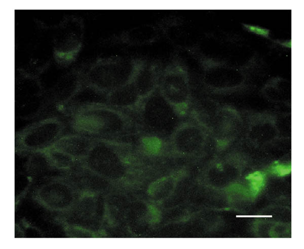
Figure 6.

Deposition of laminin by KIM-2 cells. A confluent monolayer of undifferentiated KIM-2 cells was stained with an antibody to laminin and detected by FITC-conjugated secondary antibody. Control slides with secondary antibody alone were completely blank. Scale bar 25 μm.
