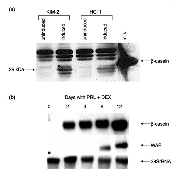
Figure 9.

Analysis of the differentiation capacity of KIM-2 and HC11 cells in response to lactogenic hormones on tissue culture plastic. (a) Endogenous β-casein expression was analyzed in KIM-2 and HC11 cells by western blot. Total cell lysates (20 μg) were prepared from KIM-2 cells or HC11 cells grown to confluence and induced with the lactogenic hormone cocktail of insulin, prolactin and dexamethasone for 4 days at 37°C. Uninduced cells were grown to confluence in growth medium. A defatted mouse milk sample was used as a positive control for the murine β-casein polyclonal antibody (diluted 1:10 000). Intracellular β-casein appeared to be a doublet at approximately 29 kDa in extracts from both cell lines, whereas a 32kDa secreted β-casein was detected in defatted milk. This size discrepancy can be accounted for by differences in phosphorylation states between intracellular and secreted forms of the protein. (b) WAP and β-casein expression were analyzed in KIM-2 cells by northern blot. Total RNA (20 μg) was prepared from KIM-2 cells grown to confluence at 37°C and harvested or induced with lactogenic hormones for the time period indicated. WAP mRNA was detected after 8 days exposure to lactogenic hormones. Figures are representative of three separate experiments, although induction of WAP expression was variable and occurred between 4 and 8 days after hormone treatment in different experiments.
