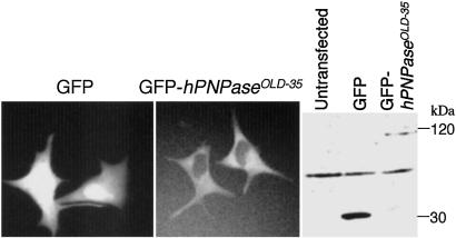
Fig 6.
Cellular localization of hPNPaseOLD-35. Cellular localization of hPNPaseOLD-35 was assessed by using a GFP-hPNPaseOLD-35 expression plasmid. (Left) HO-1 cells were transiently transfected with a GFP-hPNPaseOLD-35 construct and observed by fluorescent microscope (×400). (Right) Western blot of GFP and GFP-hPNPaseOLD-35.

