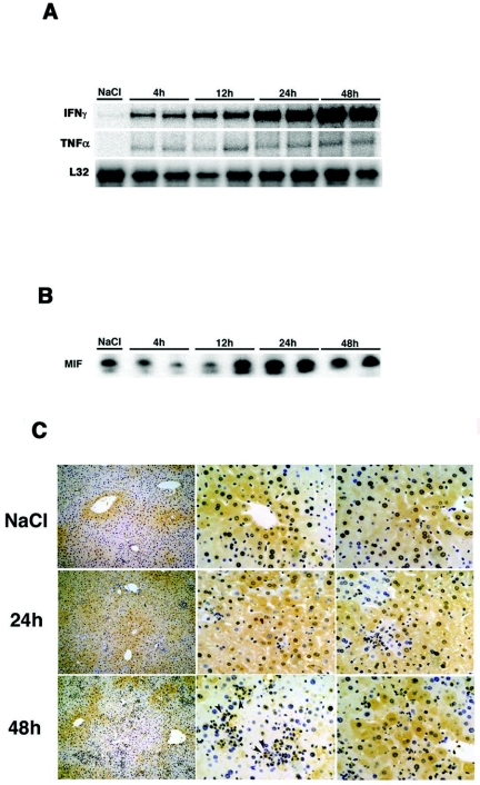
FIG. 2.
MIF expression in the liver after CTL injection. (A) Age- and sex-matched transgenic mice (lineage 107-5) were injected with 4 × 10⁶ CTLs (6C2) and sacrificed at the indicated time points. Total hepatic RNA (10 μg) was isolated from the livers at various time points and analyzed for cytokine expression by RPA. (B) Cell lysis, protein electrophoresis, and Western blotting analyses were performed as described previously (14). The membranes were probed with an anti-MIF antibody and then incubated with a horseradish peroxidase-conjugated anti-rabbit immunoglobulin G (IgG) secondary antibody. Detection was performed using an ECL system. (C) Immunohistochemical staining with a rabbit anti-mouse MIF MAb was performed using an avidin-biotin-peroxidase complex technique. Briefly, tissue slices were incubated with the anti-mouse MIF MAb overnight, followed by treatment with 3% H2O2 in absolute methanol to inhibit endogenous peroxidase activity. Next, the slices were incubated with a biotinylated secondary antibody for 10 min, washed, and incubated with an avidin-biotin-peroxidase complex reagent. Finally, the slides were treated with 0.06% diaminobenzidine and 0.01% H2O2 in 0.05 M Tris-HCl buffer (pH 7.6) for 10 min. The arrowhead indicates macrophages.

