
FIGURE 3.
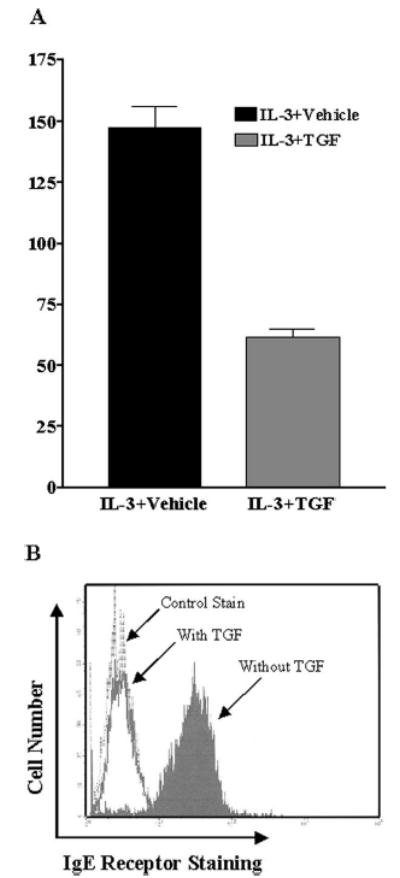
TGF-β1 inhibits FcεRI surface expression on mouse peritoneal mast cells and cultured human mast cells. A, Mouse peritoneal lavage cells were cultured in IL-3 and SCF with or without TGF-β1 for 3 days. Data shown are the average and SD of four separate populations, showing the mean fluorescence intensity of FcεRI staining on Kit-positive cells (mast cells) in the presence or the absence of TGF-β1. B, Human mast cells were cultured in SCF with or without TGF-β1 (15 ng/ml) for 3 days. FcεRI surface expression was determined by flow cytometry using anti-FcεRIα or a control Ab. Results are representative of two different mast cell cultures.