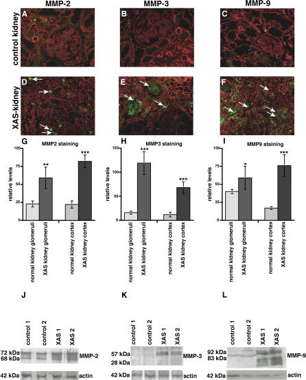
Figure 2. MMP Expression in Kidneys of Patients with XAS and in Normal Human Kidneys.
(A–F) Frozen sections of normal human kidneys and kidneys from patients with ESRD due to XAS were stained with antibodies to MMP-2, MMP-3, and MMP-9 (FITC-green), plus laminin (Rhodamine red). Representative stainings of samples obtained from a kidney designated for transplantation or from normal portions of resected kidneys with renal cell carcinoma are displayed. (A–C) Control. (D–F) Kidneys ( n = 2) obtained from male patients with XAS.
(G–I) The bar graphs summarize the quantification of MMP-2–staining (G), MMP-3–staining (H), and MMP-9–staining (I). The left bars summarize the evaluation of MMP-staining in the glomeruli; the right bars summarize the MMP-staining in the total kidney cortex in each panel. *** p < 0.001 versus control; ** p < 0.005 versus control; * p < 0.01 versus control.
(J–L) Total protein was isolated from normal human kidneys and from two kidneys obtained from two different patients with ESRD due to XAS. Protein was analyzed by SDS-PAGE and immunoblot using specific antibodies to MMP-2 (J), MMP-3 (K), and MMP-9 (L). The lower blots in each panel display the actin control blot to control for equal protein loading.

