Fig 3.
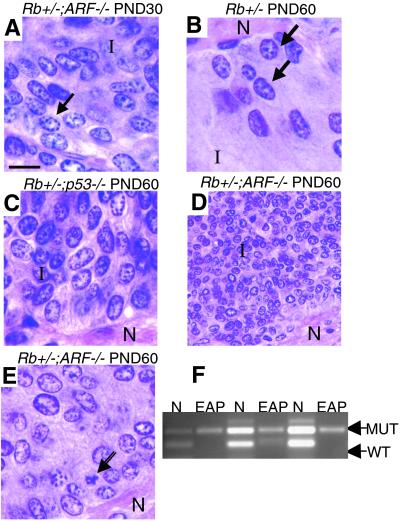
EAPs appear in the pituitaries of Rb+/−;ARF−/− compound mutants earlier than those in Rb+/− and Rb+/−; p53−/− compound mutant mice and are larger by PND 30. Comparison of hematoxylin-stained EAPs from Rb+/−, Rb+/−;ARF−/−, and Rb+/−; p53−/− compound mutant mice. (A) EAP of a PND 30 Rb+/−;ARF−/− compound mutant mouse (×1,000) showing dense groups of abnormal cells in the intermediate lobe (I) containing coarse chromatin, and large, irregular nuclei at the border with the posterior (neural) lobe. (B) EAP of a PND 60 Rb+/− mouse at the intermediate-neural lobe border (×1,000). Arrows indicate abnormal cells. Note the size of this EAP relative to the lesion in A. (C) EAP of a PND 60 Rb+/−;p53−/− compound mutant mouse showing a large lesion with densely packed abnormal cells in the intermediate lobe (×1,000). (D) Low-power view of an EAP of a PND 60 Rb+/−;ARF−/− compound mutant mouse (×400) showing typical large cluster of intermediate lobe (I) tumor cells near the neural lobe (N). (E) Higher-power (×1,000) view of one of the same lesions (D) demonstrating typical morphology of tumor cells. Note the presence of an apoptotic cell (arrow). (F) LOH at the Rb locus is observed in PND 60 EAPs from Rb+/−;ARF−/− mice. PCR analysis of DNA from laser capture-dissected samples shows retention of mutant allele with absence of WT allele in captured EAPs, while both alleles are intact in adjacent normal pituitary (N). (Calibration bar: 10 μm for A–C and E; 25 μm for D.)

