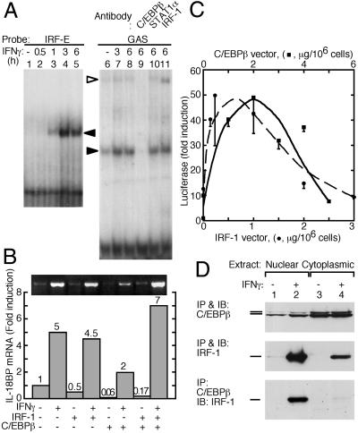
Fig 4.
Physical association and role of IRF-1 and C/EBPβ in IL-18BP gene induction. (A) EMSA of double-stranded DNA probes corresponding to bases −33 to −75 (IRF-E, lanes 1–5) and −8 to −55 (GAS, lanes 6–11). Hep G2 cells were treated with IFN-γ for the indicated times, and nuclear extracts were allowed to react with the IRF-E or GAS probes. Shifted bands are indicated by filled arrowheads. The GAS complex was also subjected to supershift with the indicated antibodies. The supershifted band is indicated by an open arrowhead. (B) Semiquantitative RT-PCR of IL-18BP mRNA from Hep G2 cells that were transfected with the indicated combinations of IRF-1 or C/EBPβ expression vectors. Where indicated, IFN-γ was added and cells were harvested 5 h later. Values were normalized to β-actin mRNA. (C) Luciferase activity in cells transfected with the luciferase reporter vector pGL3(1272), containing the complete IL-18BP promoter, together with the indicated concentrations of pCDNA3-IRF-1 (circles) and a fixed concentration of pCDNA3-C/EBPβ concentration of pCDNA3-IRF-1 (1 μg/106 cells). Alternatively, cells were transfected with the indicated concentrations ofpCDNA3-C/EBPβ (squares) and a fixed pCDNA3-IRF-1 (0.1 μg/106 cells). Luciferase activity was normalized by the β-galactosidase activity. (D) Immunoblots of nuclear and cytoplasmic extracts of cells treated with IFN-γ (100 units/ml, 2 h). Extracts were immunoprecipitated and immunoblotted with the indicated antibodies.

