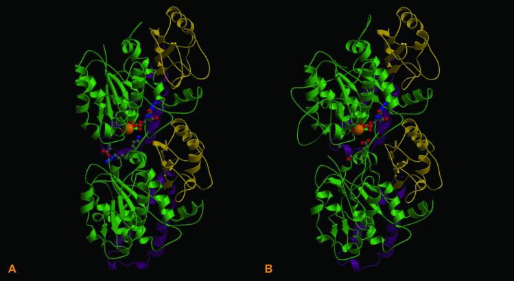
Fig 3.
Comparison of the refined crystal structure of the bovine αβ tubulin dimer (A) with the modeled P. dejongeii BtubA/BtubB structures (B). Each structure indicates the position of GTP and a Mg2+ ion at the intradimer active site (N-site). The hydrophobic C-terminal loop and helices are marked in magenta. Rossmann folds are marked in green.

