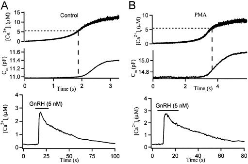
Fig 1.
PMA lowers the calcium threshold for secretion in gonadotropes. Steady UV illumination generates ramp [Ca2+]i increases with intracellular caged Ca2+ and measures [Ca2+]i by using a Ca2+ indicator, fura-6F. Simultaneous time courses of [Ca2+]i and Cm for a single control gonadotrope (A) and for a PMA-treated (for 2–3 min) gonadotrope (B). Note at the same [Ca2+]i level, Cm only begins to increase in the control cell, whereas it already takes off in the PMA-treated cell. At the end of the experiments, the cells were challenged by 5 nM GnRH to verify them as gonadotropes (Lower).

