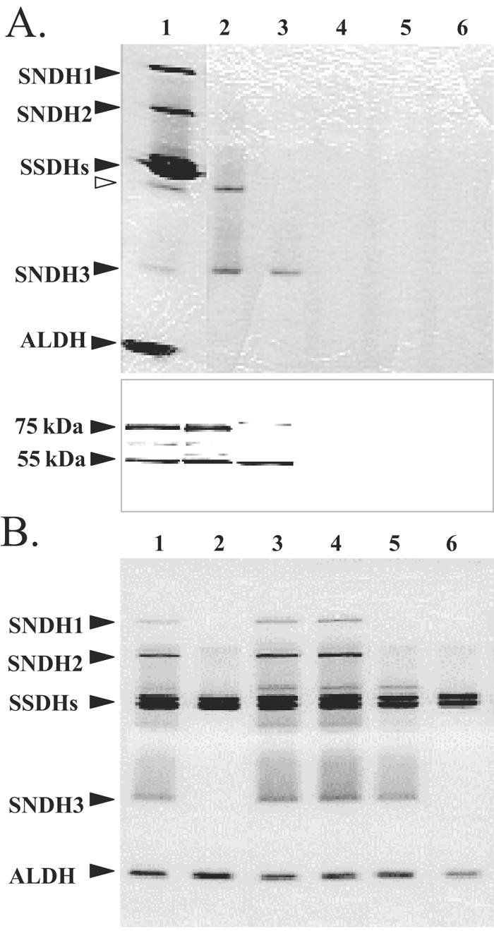
FIG. 5.

Expression of intact and truncated sndH genes in E. coli (A) and K. vulgare (B). (A) E. coli cells harboring pUCSN series plasmids: activity staining (upper gel) and Western blot analysis (lower gel) of CFE proteins. For the upper gel, CFE proteins (15 μg) were incubated with PQQ and CaCl2 for 10 min at 25°C and subjected to native PAGE (10% polyacrylamide gel), followed by activity staining using l-sorbosone. The locations of SNDH, SSDH, and ALDH proteins are indicated on the left. The open arrowhead indicates the position of the fourth SNDH band. For the lower gel, CFE proteins (5 μg) were subjected to SDS-PAGE (12.5% polyacrylamide gel) and blotted onto a polyvinylidene difluoride membrane. The protein bands were detected using rabbit polyclonal anti-SNDH antiserum. The locations of 75-kDa and 55-kDa subunits are indicated on the left. Lane 1, K. vulgare DSM 4025; lanes 2 to 6, E. coli JM109 harboring pUCSN2004 (lane 2), pUCSN2003 (lane 3), pUCSN2002 (lane 4), pUCSN2001 (lane 5), and pUC18 (lane 6). (B) K. vulgare sndH::Km cells harboring pVSN series plasmids. CFE proteins (16 μg) were subjected to native PAGE (10% polyacrylamide gel), followed by activity staining using l-sorbosone. Lane 1, K. vulgare GOMTR1; lane 2, GOMTR1SN::Km; lanes 3 to 6, GOMTR1SN::Km harboring pVSN117 (lane 3), pVSN106 (lane 4), pVSN114 (lane 5), and pVK100 (lane 6).
