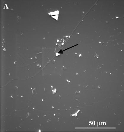
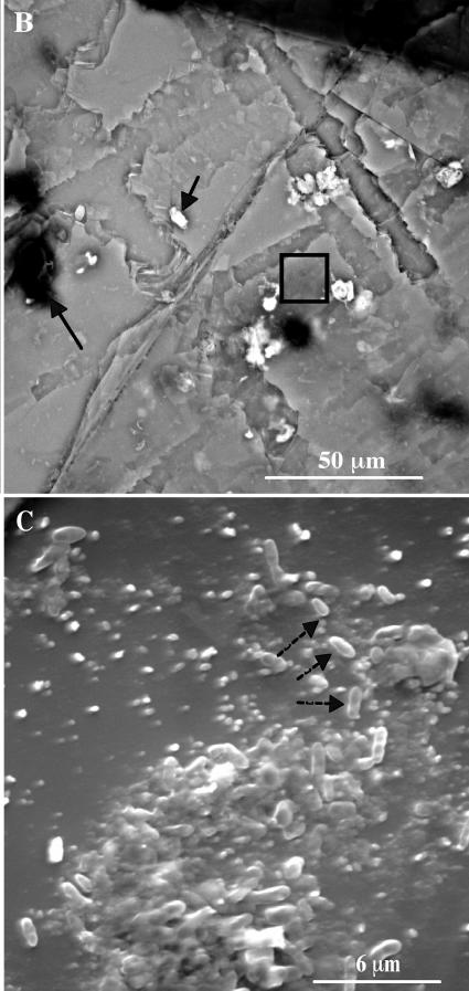
FIG. 2.
SEM photography of biotite surfaces without plant and bacteria (A) and with plant inoculated with the bacterial strain PML1(12) (B and C). The white and black areas presented with a black arrow correspond, respectively, to iron precipitates and carbon deposits. Panel C corresponds to an enlargement of the square region in panel B. The dotted arrows point to bacteria on the biotite surface.


