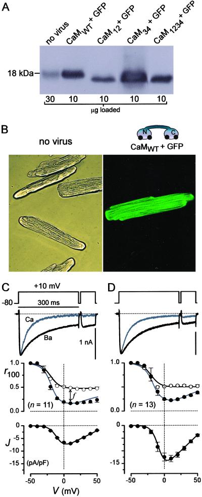
Fig 1.
Delivery of engineered CaMs to myocytes. (A) CaM Western blots from rat cardiocytes (9), cultured for 2 d after infection. Lysate amount per lane is indicated at the bottom. (B) Micrographs (×400) relating to A, with uninfected cells in bright-field (Left) and cells with AdIR-CaM under fluorescence (Right). (C and D) L-type channels in cells relating to those in B. (Top) Exemplar currents with Ca2+ (gray) or Ba2+ (black) as the charge carrier. The Ca2+ trace was amplified ≈×3 to match the Ba2+ traces; scale bars for Ba2+. (Middle) Mean CDI properties shown by Ca2+ and Ba2+ r100 curves, and the average of n cells. f is defined at +10 mV. ○, Ba2+; •, Ca2+. (Bottom) Peak current density, with Ca2+, the mean from the same n cells. GFP inexplicably enhanced the current.

