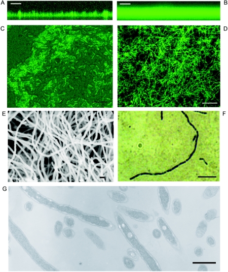
FIG. 2.
Structure of L. pneumophila biofilms. The L. pneumophila Knoxville-1 strain expressing GFP was observed under CSLM (A to D). x-z plane projection of 25°C biofilm at day 18 (A) and 37°C biofilm at day 6 (B); x-y plane projection of 25°C biofilm at day 8 (C) and 37°C biofilm at day 4 (D). The L. pneumophila Knoxville-1 biofilm at 37°C on day 4 was observed under scanning electron microscope (E), and its filamentous cell stained with HCl-Giemsa was observed under the light microscope (F). (G) A section of the filamentous cells of L. pneumophila Philadelphia-1 strain was observed under the transmission electron microscope. Bars, 50 μm (A and B), 10 μm (C and D), 1 μm (E), 5 μm (F), and 1 μm (G). Images were processed and compiled with Adobe Photoshop 7.0 software.

