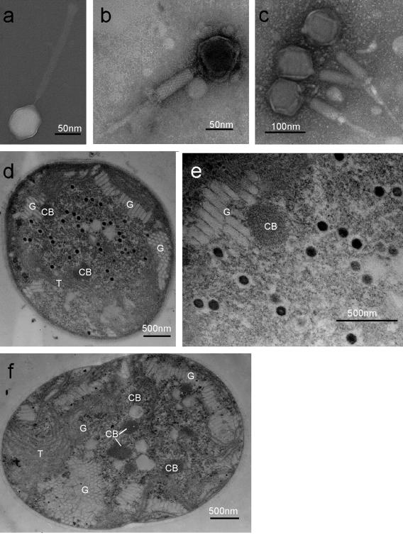
FIG. 1.
Transmission electron micrographs of cyanophage Ma-LMM01 and its host, M. aeruginosa NIES298. (a) Negatively stained virion of Ma-LMM01 with an extended tail; (b) negatively stained virion of Ma-LMM01 with a contracted tail; (c) negatively stained virions of Ma-LMM01 with a contracted tail that were purified using CsCl step gradient ultracentrifugation; (d) thin section of an M. aeruginosa cell 52 h after inoculation with Ma-LMM01; (e) higher magnification of the Ma-LMM01 particles in panel d; (f) thin section of a healthy cell of M. aeruginosa. CB, carboxysome; G, gas vesicle; T, thylakoid.

