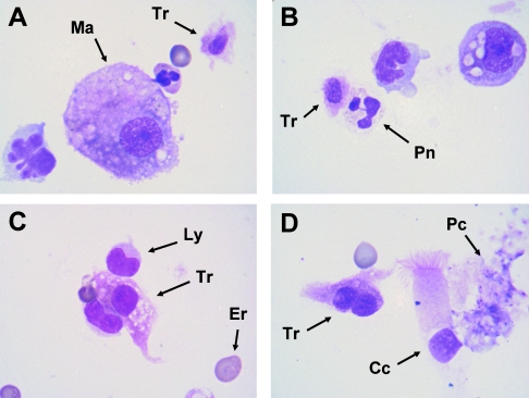
FIG. 1.
Cytological appearance of trichomonad cells in the BAL sample (May-Grünwald-Giemsa staining). (A) A trichomonad cell (Tr) with an oval nucleus is easily recognizable in the vicinity of a macrophage cell (Ma). (B) A trichomonad cell is seen in contact with a neutrophile polymorphonuclear (Pn). (C) An amoeboid trichomonad with a round nucleus is seen between two lymphocytes (Ly); an erythrocyte (Er) gives the scale (diameter, 7 μm). (D) An amoeboid trichomonad exhibiting two nuclei is seen in the vicinity of a bronchial ciliated cell (Cc) and an aggregate of Pneumocystis organisms (Pc). Magnification, ×1,000.

