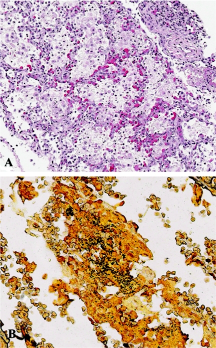
FIG. 1.
Hematoxylin and eosin (A) and special (B) staining of lung tissues from selected patients with B. pertussis infection. (A) Lung from patient 2 showing intra-alveolar infiltrates comprised predominantly of macrophages and neutrophils, accompanied by fibrin and necrotic debris. Magnification, ×100. (B) Steiner silver staining of lung from patient 3 showing abundant coccobacilli in a bronchiole. Magnification, ×158.

