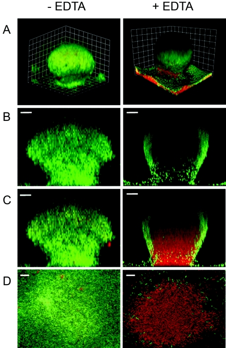
FIG. 2.
Effect of EDTA on P. aeruginosa biofilm structure. GFP-labeled P. aeruginosa biofilms were grown in flow cells for 6 days. Biofilms were grown at room temperature and treated with 50 mM EDTA for 2.5 h. The biofilm matrix and dead cells were counterstained with propidium iodide (30 μM) prior to EDTA treatment. (Left) Biofilm prior to EDTA treatment. (Right) Biofilm after EDTA treatment. The images were acquired by CSLM. (A) Three-dimensional reconstruction. The combined green (GFP) and red (propidium iodide) channels are presented. The squares are 15 μm on each side. (B) Sagittal views of the green channel only. (C) Sagittal view showing the combined red and green channels. Bars, 20 μm. (D) A 0.5-μm slice of the internal region of the biofilm. The combined red and green channels are presented. Bars, 10 μm.

