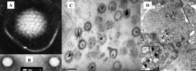
FIG. 1.
Electron micrographs of CNGV. (A and B) CNGV harvested from infected KFC was purified by sucrose gradient centrifugation, and virus was negatively stained with 2% phosphotungstate. The core size was in the 96- to 105-nm range, with an average diameter of 103 nm. Bar, 0.1 μm. (C) Thin sections were made for ultrastructural analysis by transmission electron microscopy. Purified virus pellets were fixed with 3% glutaraldehyde in 0.1 M sodium cacodylate and stained with uranyl acetate and lead citrate. (D) Ultrastructural appearance of CNGV particles in infected kidney at 8 days postinfection. This cell harbors several cytoplasmic viral particles with round electron-dense cores (magnified in the inset). Bars, 200 nm.

