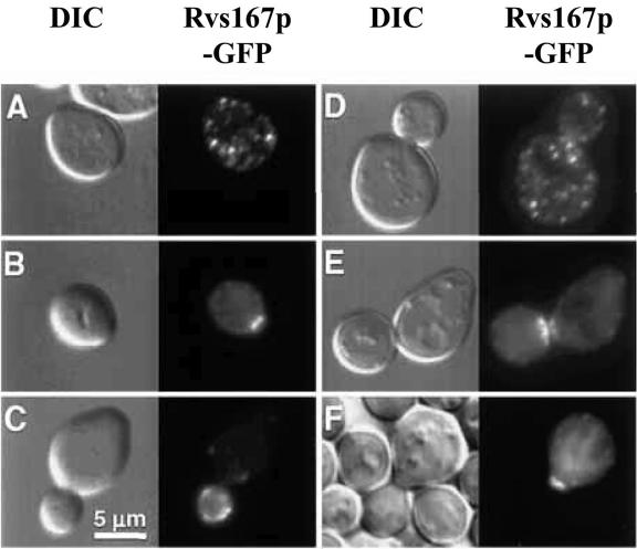
FIG. 3.
Subcellular localization of Rvs167p in growing and mating yeast cells. Shown are budding yeast cells that express a fusion protein (Rvs167p-GFP) that comprises full-length Rvs167p with GFP inserted between the BAR and GPA-rich domains of Rvs167p. The same fields of cells were viewed by fluorescence optics (right) to visualize GFP and by differential interference contrast (left) to visualize the cell profiles. A, unbudded cell; B, cell at an early stage of bud emergence; C, cell with small bud; D, cell with large bud; E, cells undergoing division (cytokinesis); F, cells arrested in G1 and forming a mating projection (shmoo) after pheromone treatment (only one cell expressing Rvs167p-GFP is depicted in this panel). In vegetatively growing cells Rvs167p-GFP localizes to large cortical patches that polarize to nascent bud sites, small buds, and the bud neck during cell division (cytokinesis). In mating cells Rvs167p-GFP concentrates at the tip of the mating projection (shmoo). (Reprinted from reference 11 with permission of the Company of Biologists Ltd.)

