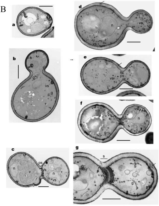
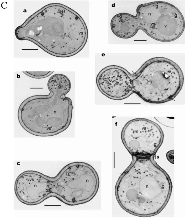
FIG.7.
Defects in vesicle fusion in rvs161 mutant cells result in vesicle accumulation. Shown are electron micrographs of wild-type (A), rvs161Δ (B), and rvs167Δ (C) yeast cells. The panels on the left depict cells grown under standard conditions,while those on the right depict cells grown in medium containing sublethal levels (3.4%, wt/vol) of Na+. Bar, 1 μm. Labeled organelles: n, nucleus; v, vacuole; vs, vesicles; g, Golgi apparatus apparatus; ps, primary septum; s, septum. The arrow indicates cell wall abnormalities. (Reproduced from reference 20 with permission of the publisher. Copyright John Wiley and Sons Ltd.)


