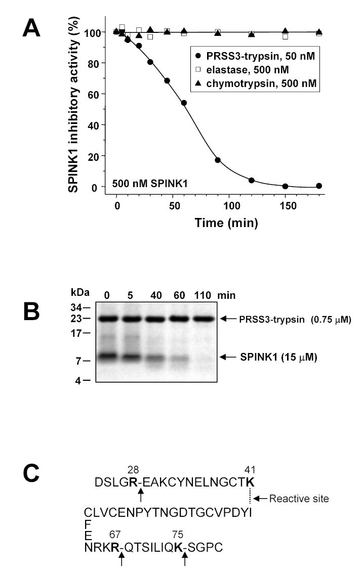
Figure 7.

Degradation of human SPINK1 by mesotrypsin. Panel A. SPINK1 (500 nM) was incubated with 50 nM mesotrypsin, 500 nM bovine chymotrypsin or 500 nM human elastase 2 (final concentrations) at 37 °C in 0.1 M Tris-HCl (pH 8.0), 1 mM CaCl2 and 1 mg/mL bovine serum albumin. Residual inhibitory activity was measured with bovine trypsin, as described in Experimental Procedures. Panel B. SPINK1 (15 μM) was incubated with 0.75 μM mesotrypsin (final concentrations) at 37 °C in 0.1 M Tris-HCl (pH 8.0) and 1 mM CaCl2. Samples were precipitated with 20 % trichloroacetic acid (final concentration) at indicated times; separated on 16 % tricine-SDS gels under reducing conditions and visualized by Coomassie blue staining. Note that SPINK1 stains very poorly, and the relative intensities of the mesotrypsin and SPINK1 bands do not reflect the actual ratio of their concentrations in the reaction mixtures. Panel C. Sites of mesotrypsin cleavage in SPINK1, as determined from N-terminal sequencing of a sample of the digestion mixture at 40 min in Fig 7B.
