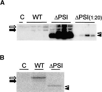
Figure 4.
Phytepsin Protein Levels Are Influenced by the Subcellular Localization.
(A) Leaf extracts from untransformed plants (C) or plants expressing wild-type Phytepsin (WT) or PhytepsinΔPSI (ΔPSI). At right, 20-fold dilutions of PhytepsinΔPSI plants were loaded to highlight the drastically increased levels compared with the wild type.
(B) Cellular protein levels detected by pulse labeling and immunoprecipitation. Protoplasts were prepared from transgenic plants used in the experiment described in (A), labeled in vivo, and immunoprecipitated with Phytepsin antiserum. Annotations are as in (A). Note that the levels observed for the truncated proteins are slightly lower than those of the wild-type protein, in sharp contrast to (A).
Processed and unprocessed forms of either molecules are indicated by arrows (WT) or arrowheads (ΔPSI) as in Figures 1 and 2.

