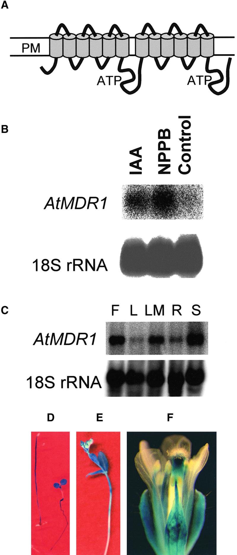
Figure 1.

Topology and Expression Pattern of AtMDR1.
(A) Two groups of six transmembrane helices are predicted, each followed by cytoplasmic domains containing ATP binding domains. An alternate model having seven membrane-spanning helices in the first group also is supported. PM, plasma membrane.
(B) NPPB- and IAA-induced expression of AtMDR1 mRNA. Each lane of the gel contained 30 μg of total RNA extracted from Arabidopsis seedlings grown for 5 days in the presence or absence of 20 μM NPPB or 1 μM IAA, and the blot was probed with 32P-labeled AtMDR1 genomic DNA and also with 18S rRNA to confirm equal RNA loading.
(C) Tissue-specific expression pattern of AtMDR1 mRNA. Each lane contained 30 μg of total RNA from flowers (F), rosette leaves (L), rosette leaves plus shoot apical meristem (LM), roots (R), and seedlings (S). A blot of the RNA gel was probed with AtMDR1 genomic DNA and also with 18S rRNA to confirm equal RNA loading.
(D) GUS activity patterns in etiolated (left) and light-grown (right) AtMDR1::GUS seedlings. Light increased AtMDR1 promoter activity in cotyledons and roots but suppressed it in hypocotyls of these 5-day-old seedlings.
(E) GUS activity in the primary inflorescence of an AtMDR1::GUS plant. Staining was strongest in the apical portions of the stem and the youngest cauline leaves.
(F) GUS activity pattern in the flower of an AtMDR1::GUS plant. All floral whorls except petals displayed staining.
