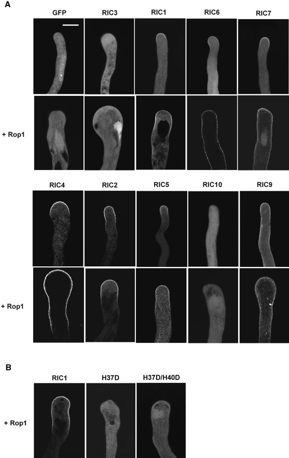
Figure 5.
Subcellular Localization of GFP-RICs with and the Effect of Rop1 Expression on Their Localization in Tobacco Pollen Tubes.
Tobacco pollen grains were bombarded with various constructs and germinated as described in Figure 4. Approximately 5 hr after bombardment, GFP localization in transformed pollen tubes was analyzed using confocal microscopy.
(A) GFP-RIC localization was analyzed either in tubes expressing GFP-RIC alone (top) or coexpressing Rop1 (bottom). All images shown are 2-μm median sections. The strong fluorescence near the tips of tubes expressing GFP-RIC3 plus Rop1 most likely represents GFP-RIC3 localization to the vegetative nucleus. Bar = 20 μm.
(B) Mutations in the CRIB motif (see Figure 1B) eliminate the localization of GFP-RIC1 to the apical PM domain.

