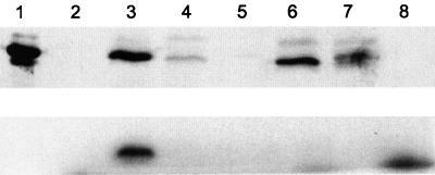
FIG. 3.
Western analysis and uridylylation of A. vinelandii GlnK and GlnKY51F in E. coli. (Top) Western blot of E. coli extracts incubated with 2-oxoglutarate and UTP for 30 min. (Bottom) Autoradiograph of the same extracts labeled with [α32-P]UTP. Lanes: 1, purified GlnKHis6 as a size standard; 2, CK1007 (glnB glnK); 3, CK1007(pPR113); 4, CK1007(pPR115); 5, CK1008 (glnB glnK glnD); 6, CK1008(pPR113); 7, CK1008(pPR115); 8, no protein.

