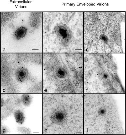
FIG. 1.
Immunoelectron microscopy of extracellular and primary enveloped virions observed in cells infected with recombinant viruses expressing VP16gpf (a to c), VP22gfp (d to f), or VP13/14yfp (g to i). Panels c, f, and i represent lower-magnification images of the primary enveloped virions shown in panels b, e, and h respectively. Scale bars, 100 nm.

