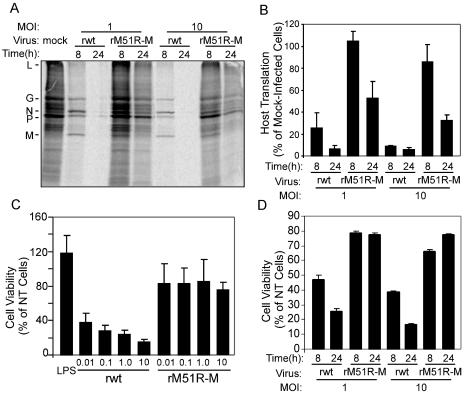
FIG. 1.
rM51R-M virus is defective at inhibiting host gene expression in mDC. mDC were infected with rwt or rM51R-M virus at a multiplicity of infection of 10 or 1 PFU/cell or were mock infected as a control. At 8 and 24 h postinfection, cells were labeled with a 15-min pulse of [35S]methionine (100 μCi/ml) and harvested. Lysates were subjected to SDS-PAGE, and labeled proteins were quantitated by phosphorimaging. (A) Representative image from analysis of rwt and rM51R-M virus-infected mDC. (B) Host protein synthesis was determined from images similar to that shown in panel A for regions of the gel devoid of viral proteins between the L and G proteins. The results are shown as percentages of the mock-infected control value and are the means ± standard errors of three independent experiments. (C) Cells were infected with the rwt and rM51R-M viruses at multiplicities of 0.01, 0.1, 1, and 10 PFU/cell or were treated with LPS, and live cells were measured by an MTT assay at 24 h postinfection. Data are expressed as percentages of the cell viability of untreated cells and are the means ± standard errors of four experiments. (D) Viability of mDC infected with rwt or rM51R-M virus at a multiplicity of 1 or 10 PFU/cell for 8 or 24 h. Data are means ± standard errors of three experiments.

