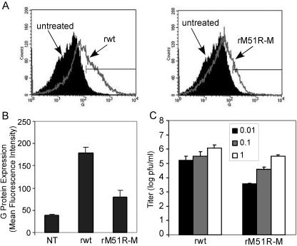
FIG. 2.
mDC are semipermissive for rwt and rM51R-M virus infections. The efficiency of G protein surface expression on mDC during VSV infection was determined by flow cytometry analysis of mDC infected with the rwt and rM51R-M viruses at a multiplicity of 1 PFU/cell for 24 h. (A) Representative histograms depicting increases in G protein expression in rwt and rM51R-M virus-infected cells. (B) The geometric mean fluorescence intensity of each sample was determined. Data are expressed in arbitrary values and are the means ± standard errors of four or five experiments. (C) Viral growth analysis in mDC. Cells were infected with rwt or rM51R-M virus at multiplicities of 0.01, 0.1, and 1 PFU/cell. At 24 h postinfection, supernatants were collected to determine the amounts of progeny virus by a plaque assay. Data are the means ± standard errors of three experiments.

