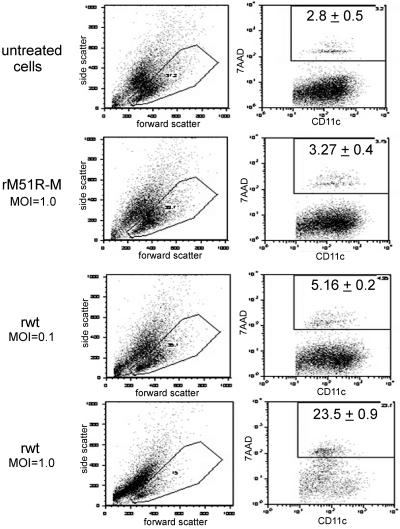
FIG. 3.
Quantitation of cell death and CD11c expression following infection with rwt and rM51R-M viruses. mDC were infected with rwt and rM51R-M viruses at multiplicities of 0.1 PFU/cell (rwt virus) and 1 PFU/cell (rwt and rM51R-M viruses). At 24 h postinfection, cells were labeled with antibodies to CD11c and incubated with 7AAD. The images shown are representative dot plots depicting the forward and side scatter of each population and CD11c versus 7AAD staining within the low-side-scatter gate. The percentage of 7AAD-positive cells in the gated CD11c+ population was determined. Data represent the means ± standard deviations of three experiments.

