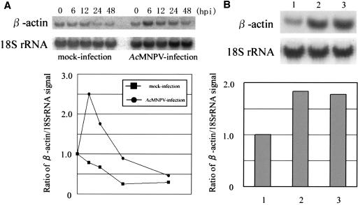
FIG. 3.
Northern hybridization assays using 32P-labeled β-actin and 18S rRNA cDNA probes. (A) Northern hybridization was performed using total RNA purified from AcMNPV-infected HeLa14 cells at the times shown in the panel. Kinetics of the expression of β-actin is presented as a sequential line graph below the autoradiograph and the values are shown as the ratio of β-actin to 18S rRNA on the basis of the signal intensities obtained from the Northern hybridization. Circles and boxes in the graph indicate the kinetics for AcMNPV-infected and mock-infected cells, respectively. (B) Northern hybridization assay using total RNA from mock-infected (1), AcMNPV-infected (MOI of 30) (2) and UV-inactivated virus-infected (3) cells. The bar graph below the autoradiograph shows the ratio of hybridization signal for β-actin to that for 18S rRNA.

