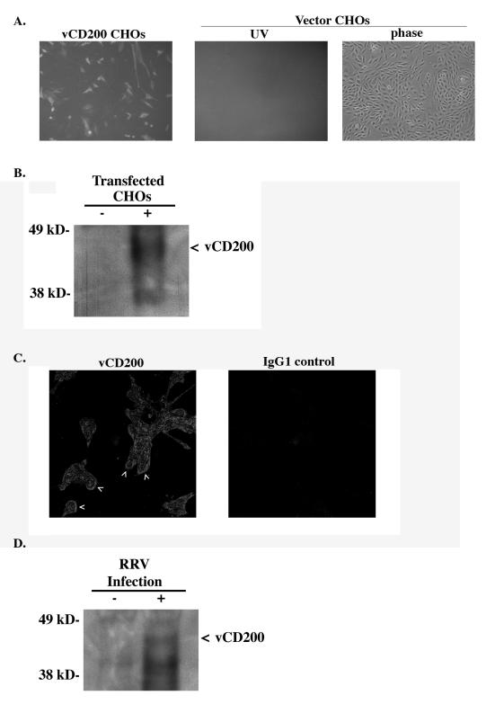
FIG. 3.
Expression analysis of vCD200. (A) vCD200 monoclonal antibodies (11B8) specifically detect the expression of vCD200 in CHO cells transfected with a full-length vCD200 expression vector and not in cells transfected with vector only. (B) Western blot analysis of vCD200 (arrowhead) from supernatants of CHO cells transfected with a FLAG-tagged vCD200 expression vector (+) or control vector (−), immunoprecipitated with an anti-FLAG antibody, and probed with antibody 11B8. (C) Specific staining of vCD200 was observed on the surfaces (arrowheads) of infected cells at 52 h postinfection. No specific staining was observed when infected cells were incubated with the isotype control antibody. Images represent an area of 281 μm by 281 μm. (D) Immunoprecipitation of vCD200 (arrowhead) from supernatants of RRV-infected fibroblasts at 72 h postinfection (+) or of uninfected cells (−).

