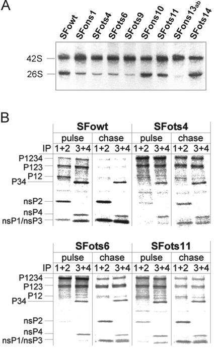
FIG. 1.
RNA and protein analysis of the recombinant virus strains after shift-up and equilibration to the restrictive temperature. (A) Viral RNAs labeled for 1 h with [3H]uridine were extracted, analyzed in agarose gels, and visualized by fluorography. Virus strains are indicated at the top, and the positions of 42S and 26S RNAs are marked. (B) Proteins were labeled for 15 min with [35S]methionine. The sample from one parallel plate was extracted immediately after the pulse, while the other plate was chased with excess cold methionine for 1 h before extraction. The samples were immunoprecipitated with combinations of antibodies against the ns proteins and analyzed by sodium dodecyl sulfate-polyacrylamide gel electrophoresis and fluorography to detect all of the polyprotein precursors and mature proteins (16). The virus strain, sample (pulse or chase), and antibody combinations used for immunoprecipitation (IP, numbers denote antibodies against the ns proteins) are indicated at the top. The positions of proteins are marked on the left. NsP1 and nsP3 migrate very similarly but can be visualized on adjacent lanes due to the antibody combinations used.

